Article: Signs of Lip Filler Migration: What You Should Know
Signs of Lip Filler Migration: What You Should Know
You notice a faint ridge above your upper lip while applying makeup. It wasn't there immediately after your appointment, or perhaps it was hidden by swelling, but now the border looks blurred and one side feels firmer than the other. Searching for answers can quickly turn a normal healing response into a frightening diagnosis.
The signs of lip filler migration deserve careful assessment, not panic. A lump, fullness, or asymmetry may result from swelling, misplaced product, infection, inflammation, or a foreign-body reaction. Migration is only one possibility, and appearance alone rarely tells the whole story.
Understanding Lip Filler Migration
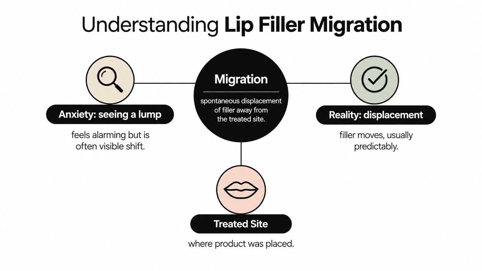
Lip filler migration means injected material has moved spontaneously away from its original treatment site. In practice, this differs from the temporary tissue response many patients experience during the first days after treatment. Swelling, tenderness, small bumps, and uneven fullness can reflect healing rather than displacement.
The defining concern is the product's location. Filler that remains within the intended injection plane may create expected fullness. Material appearing in nearby, untreated tissue can indicate migration, although examination is needed before drawing that conclusion.
A 2023 review of published lip-filler complication cases examined 53 reported cases and identified 82 total complications. Nine complications, approximately 11.0%, were classified as migration, while nodules were the most frequently documented complication overall (2023 review of lip-filler complications). These figures describe complications in a selected group of published reports, not the likelihood of migration among everyone who receives filler.
The distinction matters because lips are small, mobile structures, and several conditions can produce similar changes. People comparing lip filler types and benefits should also consider expected effects, product differences, and potential side effects before treatment.
Clinical rule: A visible change warrants assessment, but it does not prove that filler has migrated.
Assessment should compare the current contour with the original injection sites, establish when the change began, and include direct examination of the tissue. Ultrasound can help locate filler beneath the skin when the surface appearance is unclear, an often-overlooked step in consumer discussions. The BotoxBarb guide to whether fillers migrate offers patient-focused background, while persistent or progressive findings require individualized evaluation by a qualified clinician.
Identifying Clinical and Visual Signs
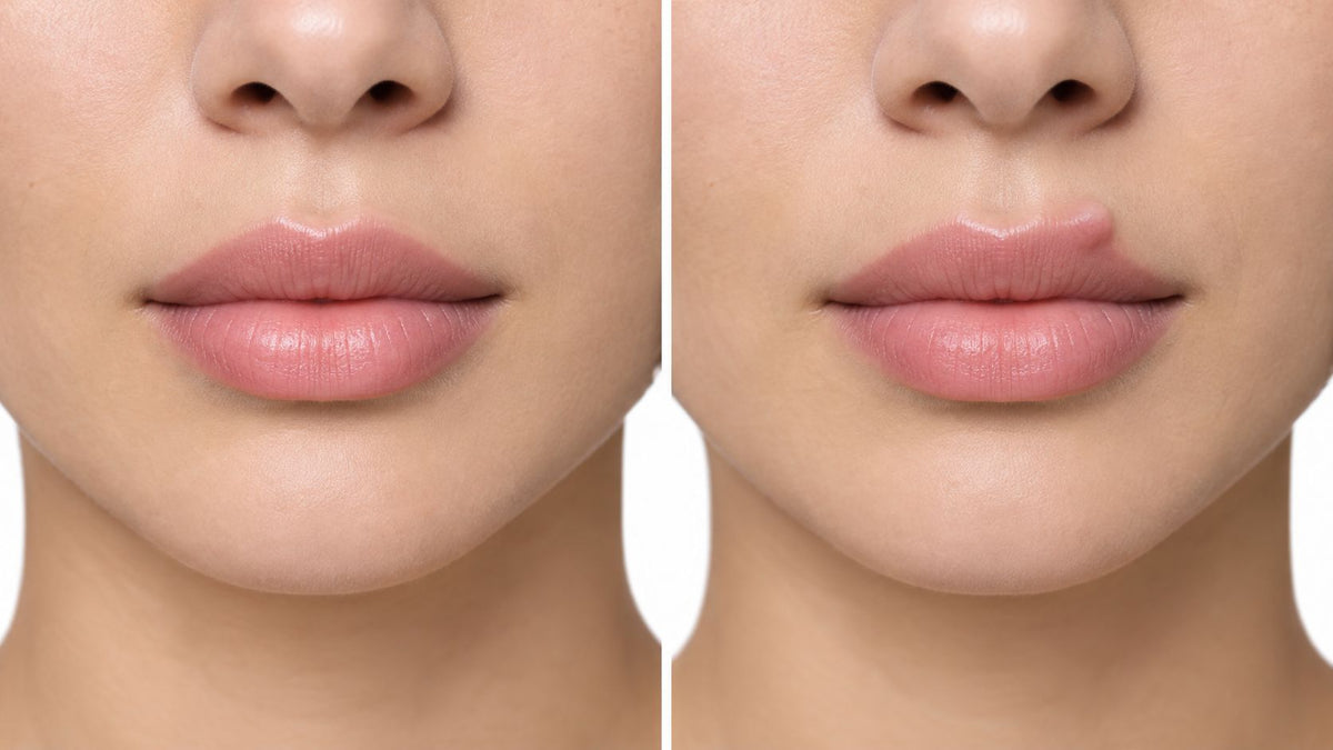
A new ridge appears above the colored lip, or a firm lump develops beneath the nose after the initial treatment period. These findings deserve assessment, especially when they sit outside the original injection area. The vermilion border marks the transition between the colored lip and surrounding skin. Filler above that boundary can create a shelf-like contour or blur the lip outline.
People may notice a localized upper-lip projection, firmness when lightly touching the skin, or fullness in tissue that was not treated. Persistent asymmetry after the expected healing period is another reason to seek review. An isolated nodule differs from broad, generalized puffiness and should be documented rather than assumed to be migration.
The FDA classifies dermal fillers as medical devices and reports that complications may occur immediately or later, including nodules, infection, inflammation, and other tissue reactions (FDA information on dermal fillers). A lump alone cannot establish migration. Its position, behavior, and associated symptoms matter more than appearance in a photograph.
What an assessment should document
A clinician should assess the full pattern:
- Location: Is the fullness outside the original injection area?
- Timing: Did it appear immediately, or after the initial healing period?
- Texture: Is it soft, firm, mobile, fluctuant, or fixed?
- Symptoms: Are tenderness, warmth, redness, or persistent swelling present?
- Symmetry: Does the same change appear on both sides?
- Movement: Does the contour change when you smile, speak, or purse your lips?
The examination should also record whether the product was injected superficially, whether older filler remained, and whether the area has been treated repeatedly. These details can support migration, but similar findings may reflect edema, infection, a granulomatous response, or product placed too close to the surface. If the physical examination cannot identify the material's location, ultrasound can help distinguish superficial filler from other causes.
Location, not the mere existence of a lump, drives the differential, and the same logic applies in other facial regions, as this discussion of cheek filler migration shows.
Distinguishing Migration from Normal Swelling
The timing and distribution of a change often provide the first useful separation. Early, diffuse swelling around the injection sites can be part of the normal inflammatory response. It may affect both lips, feel tight, and change from day to day.
Migration tends to raise more concern when the finding is localized, persistent, displaced, or progressive. Even then, it remains a clinical possibility rather than a guaranteed diagnosis. Delayed filler reactions can include erythema, edema, inflammation, nodules, and infection, and expert consensus commonly describes delayed-onset reactions as occurring more than four weeks after treatment (consensus discussion of delayed filler reactions).
| Feature | Normal Swelling | Migration |
|---|---|---|
| Distribution | Often diffuse and close to injection sites | More localized or outside the intended treatment area |
| Texture | May feel soft, tight, or generally puffy | Can feel like a persistent ridge, firm lump, or displaced fullness |
| Pattern | Often affects the treated area broadly | May appear above the vermilion border, in the philtrum, or nearby tissue |
| Progression | Usually changes as the tissues settle | May persist or become more noticeable over time |
| Diagnosis | Often supported by the expected healing pattern | Requires examination and may require imaging |
A nodule introduces another layer of uncertainty. A late, firm nodule that fails to respond to initial treatment may represent a foreign-body granulomatous reaction rather than simple product movement. Infection can also produce swelling and tenderness, while superficial placement can create visible fullness without spontaneous migration.
Practical observation: Migration is a diagnosis of location, not appearance alone.
Don't massage, puncture, squeeze, or attempt to dissolve a lump based on an online image. If bruising is part of your concern, a detailed guide to lip filler bruising can help you understand why discoloration may occur, while this explanation of swelling after filler covers common post-treatment changes. Persistent or late findings should still be examined by a qualified clinician.
Case Studies and Real-World Examples
A patient may notice a new change months after treatment, with no pain to signal a problem. In one report from the case review cited earlier, a patient developed a painless swelling six months after treatment. The material later migrated into the upper-lip mucosa, where it formed a 1.5-centimeter mass (published lip-filler complication review).
The timing and location mattered. Delayed, localized swelling differs from the broader puffiness expected soon after injection. A patient might overlook it because there was no tenderness or redness, or assume the filler had remained in its original position. Examination should establish whether the finding lies within the treated tissue and whether its texture and depth fit that explanation.

A separate report described white nodules in the mandibular sulcus that had migrated from the lower lip. The material had become visible or palpable away from the area the patient remembered being treated. This pattern makes the treatment history particularly useful, including the original product, injection sites, timing, and previous procedures.
The review recorded hyaluronic-acid-related complications among the published cases, including patients treated exclusively in the lips (case review details). Those figures describe a selected case set, not the risk for every patient or product.
Both cases share one feature: the presenting sign appeared in tissue the patient did not associate with treatment. A complete injection history is therefore part of every assessment, alongside examination and, where the findings remain uncertain, ultrasound to locate filler beneath the skin.
Causes and Risk Factors
Lip filler migration usually reflects several interacting factors, including injection depth, anatomy, movement, pressure, and product volume. A visible ridge or fullness above the border does not identify the cause by itself. Clinical examination, and ultrasound when the tissue plane is unclear, can help distinguish migrated product from superficial placement, swelling, or another tissue change.
Improper placement increases concern when filler is deposited in an unsuitable plane or too close to the surface. Normal facial movement may then make the contour more prominent or allow product to extend through nearby tissue.
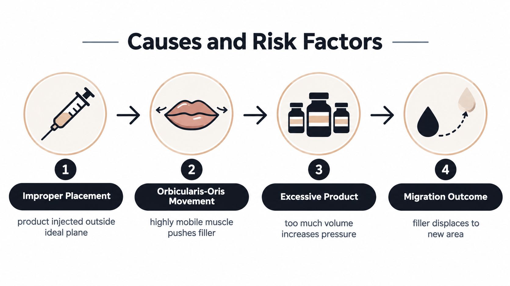
The lips move continuously during speaking, eating, smiling, and pursing. The orbicularis-oris region is therefore exposed to repeated contraction. Clinical commentary discusses placement, facial movement, and external pressure as factors that may facilitate filler displacement through surrounding tissue (clinical commentary on filler displacement).
Factors clinicians consider
- Injection plane: An unsuitable layer may leave product more visible or less stable.
- Movement: Repeated contraction around the mouth creates mechanical forces in a mobile area.
- Pressure: External pressure may contribute to movement through adjacent tissue.
- Volume: Excess filler can make an upper-lip shelf or overfilled border more apparent, depending on anatomy and product properties.
- Device choice: The FDA has warned that needle-free filler injectors use high pressure and offer insufficient control over placement. Serious injuries, including permanent harm to the skin, lips, or eyes, have occurred with these devices.
Prevention begins before injection. The provider should assess lip anatomy, identify existing filler, choose an appropriate product and plane, and avoid adding material to an area that already appears overfilled. Conservative planning reduces the risk of chasing symmetry with repeated treatment. When a contour remains unexplained, ultrasound can help locate filler before further injections or dissolving are considered.
The Role of Ultrasound in Diagnosis
Photographs can document a contour, but they can't reliably show where filler sits. A clinician may see a ridge above the lip and still need to distinguish migrated product from a superficial deposit, scar tissue, granuloma, edema, infection, or an unrelated soft-tissue mass.
High-frequency ultrasound can help locate filler, estimate its size, identify the tissue plane, and guide treatment. It may also help a clinician decide whether hyaluronidase or another intervention is appropriate, instead of treating blindly.
A 2026 ultrasound study of 126 participants found that the lip's subcutaneous layer averaged less than 1 millimeter, meaning filler was often deposited intramuscularly. The study reported spread beyond the intended site in 52% of treated patients, with a higher rate after vertical injection techniques, 73% compared with 24% for horizontal techniques (2026 ultrasound study of lip filler placement).
Those findings don't mean every ultrasound-detected spread is a clinical complication. They show that the lip's anatomy and injection technique can make product distribution more complex than a simple superficial pocket.
Why this matters: Repeated blind dissolving may remove correctly placed hyaluronic acid while missing material in a deeper or unexpected tissue plane.
Ultrasound isn't necessary for every uncomplicated post-injection change. It becomes particularly valuable when the diagnosis is uncertain, the finding is delayed, the tissue feels deep or fixed, prior treatment has accumulated, or an initial intervention hasn't produced the expected result. The examination should guide the decision to image.
Treatment and Prevention Strategies
Treatment should begin with identification, not correction. If a clinician confirms migrated hyaluronic-acid filler, hyaluronidase may be considered because it breaks down hyaluronic acid. The enzyme won't address every cause of a lump, and it doesn't treat non-hyaluronic-acid products in the same way.
A provider should first determine whether the concern is displaced product, swelling, infection, inflammation, a granuloma, or another mass. Ultrasound may improve precision when the material's location isn't clear. Dissolving the wrong structure can leave the original problem untreated and may remove filler that was correctly placed.
What helps
- Professional examination: Bring the treatment date, product information, injection locations, and photographs from before and after treatment.
- Targeted imaging: Consider high-frequency ultrasound when the tissue plane or diagnosis is uncertain.
- Individualized treatment: Use hyaluronidase only when the clinician believes hyaluronic acid is contributing to the problem.
- Conservative retreatment: Reassess the anatomy after the issue has been evaluated rather than immediately adding more filler.
- Qualified injection care: Choose a medically trained provider who understands facial anatomy, product selection, injection depth, and complication management.
What doesn't help
Don't press firmly on a persistent lump or try to puncture it. Don't use heat, light, massage, or an at-home device as a substitute for examination when swelling is unexplained. A cosmetic device can support skin-care routines, but it can't relocate or dissolve filler.
Prevention depends on thoughtful planning. The provider should account for the mobility of the lips, the existing volume, the product characteristics, and the patient's treatment history. The goal isn't maximum fullness. It's a result that remains proportionate and anatomically controlled.
When to Seek Professional Help and Next Steps
Contact your injector or another qualified medical provider for a persistent ridge, firm lump, new asymmetry, fullness outside the treated area, or any delayed change that doesn't fit the expected healing pattern. Seek urgent evaluation for rapidly increasing pain, marked redness, warmth, skin discoloration, fever, or visual symptoms.
Until you've received medical advice, avoid massaging, puncturing, squeezing, applying heat, or using light over unexplained post-injection swelling. A clinician may recommend observation, examination, ultrasound, medication, hyaluronidase, or another treatment depending on the findings.
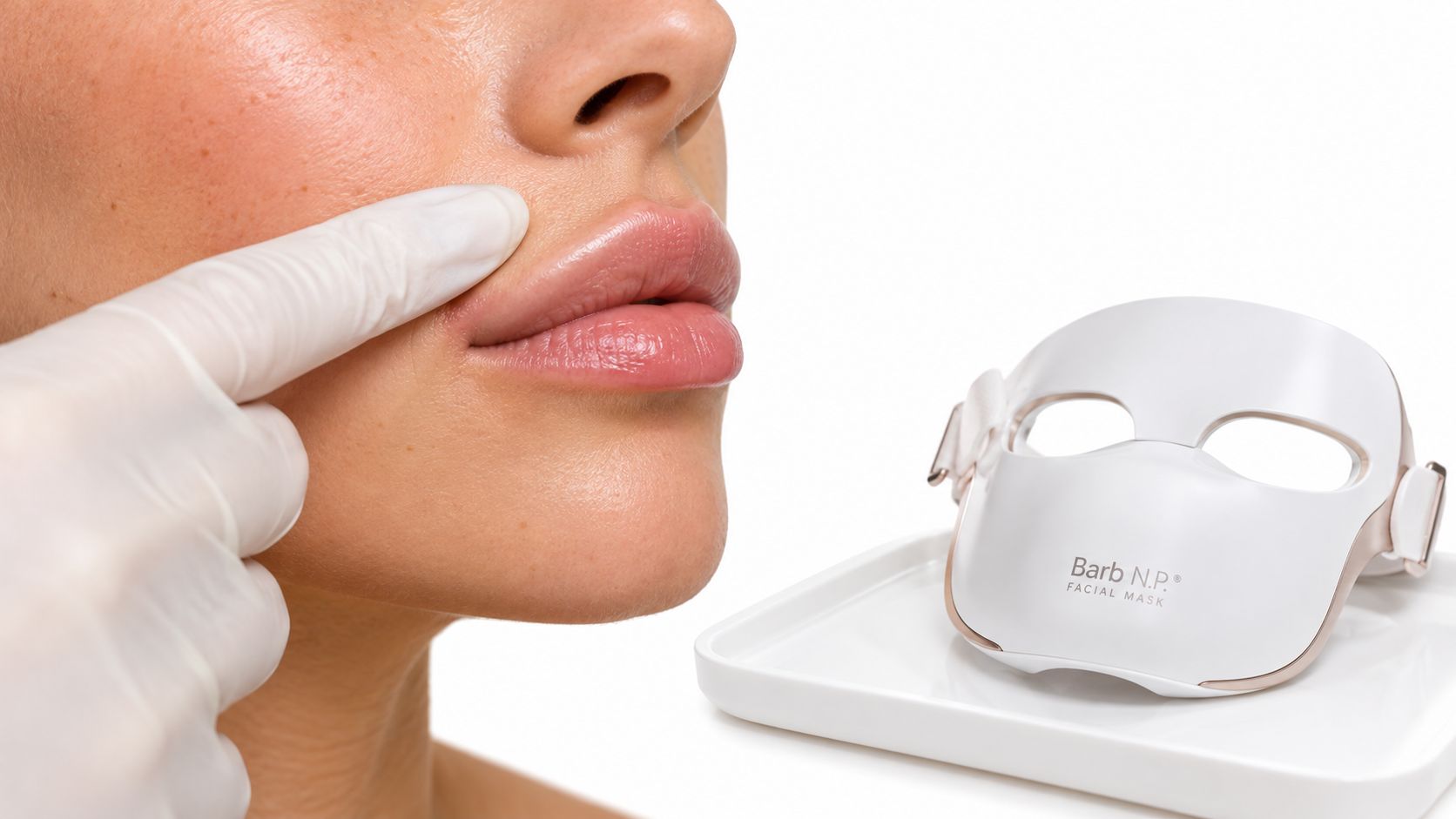
For general skin-care support, the BARB N.P. Facial Mask is a separate cosmetic wellness option. Its wireless design supports hands-free use, its face-contoured construction is intended to improve comfort, and it offers three lighting settings for different treatment goals. LED light doesn't remove, reposition, or dissolve migrated filler, and you should follow the manufacturer's instructions rather than applying a device over unexplained swelling without professional guidance.
BotoxBarb offers aesthetic consultations, injectables, LED light therapy, and curated skincare products with guidance centered on safe, individualized care. If you're concerned about filler migration or want a suitable skin-wellness device, visit BotoxBarb to explore available services and products.